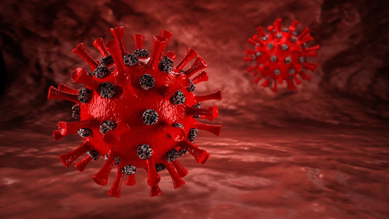
通过冷冻电子层析成像跟踪病毒病原体

通过冷冻电子层析成像跟踪病毒病原体
填写下面的表格,我们将向您发送PDF版本的电子邮件“用冷冻电子层析成像跟踪病毒病原体”

COVID-19的大流行已经显示了整个世界的重要病毒研究。事实证明,多年的研究SARS和其他冠状病毒是宝贵的基础,使研究人员能够确定SARS-COV-2尖峰蛋白的结构,并迅速开发挽救无数生命的疫苗。
In many ways, the field of virology is experiencing a renaissance, with numerous fundamental techniques having rapidly evolved over the past ten years. A significant contributor to this, other than the growth catalyzed by a global pandemic, is the standardization and commercialization of cryogenic sample preservation and analysis. In this article, we will discuss the integration of cryo-electron microscopy (cryo-EM) into virology research and the unique challenges posed by working with hazardous virus species in biosafety laboratories.
早期病毒分析
Historically, cell and virus samples have been preserved through chemical fixation or crystallization. Where whole virus analysis was impractical or impossible, the sample was broken down into key proteins for in-depth structural analysis. While these techniques have provided valuable insights into the structure, behavior and function of viruses, they are divorced from the native context in which the virus acts. And while this was acceptable for isolated, mature virus particles, it left us with gaps in our understanding of virus-cell interactions, particularly once the virus has been internalized into the cell.
用于病毒复制的细胞机械的重编程是一个复杂且高度瞬态的过程。即使通过化学固定可以进行细胞内成像,捕获这些短暂的事件也是一项挑战,尤其是有足够的细节来否定正在发生的事情。
冷冻电子显微镜
在低温样品制备中,水样标本迅速冷冻,以将其保存在无定形(玻璃体)冰中。与形成随着形成的结晶冰不同,玻璃冰分子通常保留在其液相位置,从而保留样品的周围分子结构。从本质上讲,这在近乎本地的状态下采用了标本的“快照”。
For virus analysis, cryo-preservation has been instrumental in the development of a range of electron microscopy techniques, broadly described as cryo-EM. The most common of these is single particle analysis, whose developers were awarded the2017年诺贝尔化学奖。单个粒子分析使用平均值从数百个2D透射电子显微镜(TEM)的样品的图像中产生3D分子结构,并在冰中随机定向。它通常被认为是X射线晶体学的免费,因为它可以很容易地生成蛋白质和复合物结构的结构,这些结构具有挑战性。
While single particle analysis has filled a vital niche in the world of structural analysis, it is still performed on fragmented systems isolated from their physiological context. Luckily, this need for原位观察可以通过另一种互补的低温EM技术,冷冻电子层析成像(Cryo-ET)来容纳。
冷冻电子层析成像
In electron tomography, a thin sample is progressively tilted and imaged inside a TEM. These images represent different cross-sections of the sample, which can be combined into a single high-resolution 3D representation of the specimen. Whereas single particle analysis relies on many copies of the same target molecule to capture its various orientations, tomography must utilize a precision stage that reliably tilts a single sample at discreet increments instead.
值得注意的是,这意味着可以在整个低温保存的细胞的一部分上进行冷冻-ET。虽然早期的低温ET实验仅限于固有的薄样品(或较厚的样品的薄区域),但各种冷冻粉状技术的出现使“窗户”可以直接切入冷冻细胞中,以凝视其细胞内机械。
对于病毒学,这是一个独特的机会,可以在进入细胞胞质后捕获红手的难以捉摸的病毒。特别是,冷冻-ET已被用来研究细胞内冠状病毒复制的行为。在莱顿大学医学中心领导的合作中研究人员能够可视化病毒诱导的双膜囊泡的形成,这些囊泡用于掩盖病毒RNA复制。这包括可能导致RNA转运的囊泡表面上的孔结构,将其标记为前瞻性药物靶标。


冷冻电子层析成像揭示了脊髓灰质炎病毒复制的新特征。
脊髓灰质炎病毒感染的HELA细胞的断层图3D可视化。空的衣壳:粉红色,充满RNA的衣壳:红色,蛋白质复合物将衣壳绑在膜上:黄色,腔内密度:橙色和与病毒体共包装的密集颗粒:蓝色。图像由Umeå大学Selma Dahmane提供。
生物安全实验室 - 与敌人合作
Cryo-ET of pathogenic whole viruses, and virus-cell interactions, is naturally of great interest to researchers due to the clear and present danger that viral pathogens pose to global human health.
However, biosafety laboratories have defined discrete levels of hazard that various virus samples present. Work on pathogens that pose a risk for disease requires a higher biosafety containment facility unless work is limited to isolated proteins or inactivated samples. To ensure the safety of everyone working in these facilities, researchers must take specific precautions and adhere to special protocols, including distinct instrument requirements (such as set levels of decontamination). As a result, high-end cryo-EM instruments are not yet commonly found in these higher biosafety level facilities.
然而,一些实验室在探索致病物种时表现出了令人鼓舞的结果。例如,Umeå大学的卡尔森研究小组已使用冷冻-ET研究脊髓灰质炎病毒作为模型肠病毒。在他们的工作中,他们能够捕获其宿主细胞中病毒的大部分复制周期。他们观察了病毒转化细胞内环境以促进病毒复制的方式。通过可视化这些离散机制,他们还能够看到细胞防御机制和病毒组装之间的相互作用。例如,尽管细胞触发会破坏它们,但自然自噬因素的抑制使该病毒仍能继续复制。这项工作只能归功于Cryo-Et,它开辟了令人兴奋的途径,不仅是在治疗发展方面,而且在我们对宿主病原体相互作用的基本理解中。
“想象一下,从与宿主接触的病毒接触后,一直到细胞结合,一直通过复制和释放。仅仅十年前,这可能被认为是一个幻想的梦,但是我们正在稳步接近对病毒行为的更全面的理解。” - 塞尔玛·达曼(Selma Dahmane) Umeå University
Cryo-ET opens the door to new insights into both "known" viruses whose structures might have already been determined, as well as the plethora of viruses that have yet to be studied. Many questions about their life cycles and interactions with host cells remain unanswered. Next to the determination of virus structure itself, cryo-EM, including cryo-ET, enables a close look at the virus life cycle within a host cell, e.g., entry, replication, assembly and release. There is also much to learn about how cells respond and defend themselves against viral infection. Likewise, studying the effects of drugs or drug candidates on virus life cycles and cellular defenses could better prepare us against the next major virus.
观点
病毒学有望进入一个新的发现时代,并通过冷冻保护和分析实现了近亲状态的观察。尤其是冷冻电子层析成像,可以提供洞察力,使我们目前对分子结构的了解和细胞相互作用。在更高层的生物安全设施中安装冷冻电子显微镜将使研究更广泛的病毒物种。通过对病毒作用的全面,多尺度的了解,我们可以设计更好,更有针对性,更有效的治疗方法。访问此信息不会没有挑战,但是实验者和仪器开发人员的共同努力肯定会推动我们前进到这一激动人心的未来。
About the authors
亚历克斯·伊利切夫(Alex Ilitchev)是主要科学编辑,材料和结构分析,在Thermo Fisher Scientific。
Kristian Wadel是Thermo Fisher Scientific的材料和结构分析产品营销经理。


